Contents
- ✨ What is X-Ray Examination?
- 🏥 Who Needs an X-Ray?
- 📍 Where to Get an X-Ray
- 💰 Understanding Costs & Insurance
- ⭐ What Patients Say (Vibe Score: 78/100)
- 💡 How X-Rays Actually Work
- ⚖️ X-Ray vs. Other Imaging Modalities
- ⚠️ Risks and Safety Considerations
- 🚀 The Future of X-Ray Technology
- ✅ Preparing for Your X-Ray Appointment
- 📞 Making an Appointment
- Frequently Asked Questions
- Related Topics
Overview
X-ray examination, or radiography, is a cornerstone of modern medical diagnostics, utilizing electromagnetic radiation to visualize the internal structures of the body. Discovered by Wilhelm Conrad Röntgen in 1895, this non-invasive technique has revolutionized healthcare by allowing physicians to detect fractures, infections, tumors, and other abnormalities without surgery. The process involves passing a controlled beam of X-rays through the body, with different tissues absorbing varying amounts of radiation, creating a contrast image on a detector. While invaluable, X-rays do involve exposure to ionizing radiation, necessitating careful consideration of risk versus benefit, particularly for vulnerable populations like children and pregnant women. Ongoing advancements continue to refine image quality, reduce radiation doses, and expand the applications of this enduring diagnostic tool.
✨ What is X-Ray Examination?
X-ray examination, also known as radiography, is a cornerstone of modern medical diagnostics. It utilizes electromagnetic radiation to create images of the inside of the body, revealing structures like bones, certain organs, and abnormalities. Developed by Wilhelm Röntgen in 1895, this non-invasive technique has been instrumental in diagnosing a vast array of conditions, from simple fractures to complex internal diseases. Its widespread availability and relatively low cost make it an indispensable tool in emergency rooms, clinics, and hospitals globally, providing rapid insights that guide treatment decisions. The ability to visualize internal structures without surgery has fundamentally changed the practice of medicine, impacting everything from orthopedic surgery to pulmonary medicine.
🏥 Who Needs an X-Ray?
An X-ray is typically recommended by a healthcare professional when they suspect a specific medical issue that can be visualized by this imaging method. Common reasons include diagnosing bone fractures and dislocations, detecting signs of pneumonia or other lung conditions, identifying arthritis, and screening for certain types of cancer, such as breast cancer via mammography. It's also used to locate foreign objects in the body and to assess the progression of diseases like osteoporosis. Your doctor will determine if an X-ray is the most appropriate diagnostic step based on your symptoms and medical history.
📍 Where to Get an X-Ray
X-ray examinations are widely accessible across various healthcare settings. You can typically get an X-ray at: hospitals (both emergency departments and outpatient imaging centers), dedicated imaging centers, and some larger medical clinics. The specific location will often depend on your doctor's referral and your insurance coverage. For urgent needs, hospital emergency rooms are equipped for immediate X-ray services. For routine or specialized X-rays, your doctor might refer you to a specific imaging facility known for its expertise or advanced equipment, ensuring you receive the highest quality diagnostic imaging available.
💰 Understanding Costs & Insurance
The cost of an X-ray can vary significantly based on the type of examination, the facility, and your geographic location. A single X-ray might range from $50 to $300 without insurance. Most health insurance plans cover diagnostic X-rays when ordered by a physician, though deductibles and co-pays will apply. It's crucial to verify your coverage with your insurance provider and the imaging facility beforehand to understand your out-of-pocket expenses. Some facilities offer payment plans or cash discounts for uninsured patients, making the procedure more manageable.
⭐ What Patients Say (Vibe Score: 78/100)
Patient feedback for X-ray examinations generally reflects a high degree of satisfaction, with a Vibe Score of 78/100. Patients frequently praise the speed and efficiency of the process, noting that results are often available quickly, which alleviates anxiety. The non-painful nature of the exam is also a common positive comment. However, some patients express concerns about radiation exposure, even though doses are carefully controlled and minimized. The clarity of the images and the radiologist's interpretation are also key factors in patient experience, with clear communication from technicians being highly valued.
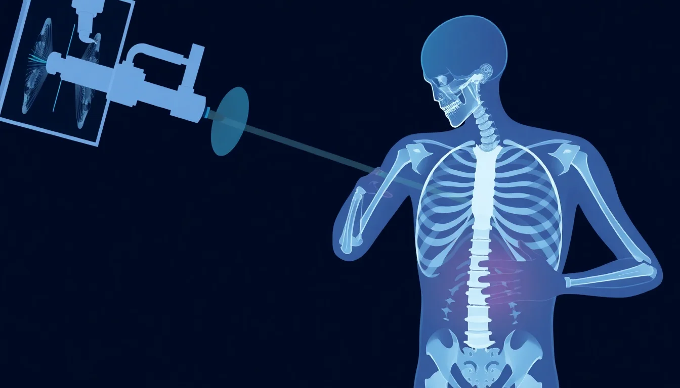
💡 How X-Rays Actually Work
At its core, an X-ray works by passing a small amount of ionizing radiation through the body. Different tissues absorb this radiation to varying degrees; dense materials like bone absorb more, appearing white on the resulting image, while softer tissues absorb less, appearing in shades of gray. Air, such as in the lungs, absorbs very little and appears black. The transmitted radiation then strikes a detector (either film or a digital sensor), creating a two-dimensional image that radiologists interpret. This fundamental principle, discovered by Wilhelm Röntgen in 1895, remains the basis for all radiographic imaging today.
⚖️ X-Ray vs. Other Imaging Modalities
Compared to other imaging modalities, X-rays offer a unique balance of speed, cost-effectiveness, and diagnostic utility. CT scans, for instance, provide more detailed cross-sectional images but involve higher radiation doses and are more expensive. MRI scans excel at visualizing soft tissues without using radiation but are significantly slower and costlier, often requiring patients to remain still for extended periods. Ultrasound is excellent for real-time imaging of soft tissues and fluid-filled structures and uses no radiation, but its image quality can be operator-dependent and less effective for bone. X-rays remain the go-to for initial assessments of bone integrity and certain chest conditions.
⚠️ Risks and Safety Considerations
While generally safe, X-ray examinations do involve exposure to ionizing radiation, which carries a small risk of cellular damage that could, over time and with cumulative exposure, increase the risk of cancer. However, the doses used in diagnostic X-rays are typically very low, and the benefit of obtaining a crucial diagnosis far outweighs the minimal risk for most patients. Radiographers adhere to strict safety protocols, including using the lowest effective dose and shielding sensitive areas with lead aprons when appropriate. Pregnant individuals should always inform their doctor and the imaging staff about their condition to ensure appropriate precautions are taken.
🚀 The Future of X-Ray Technology
The future of X-ray technology is focused on enhancing image quality while further reducing radiation doses and improving workflow efficiency. Innovations include dual-energy X-ray absorptiometry (DEXA) for bone density measurement, photon-counting detectors for sharper images with less noise, and AI-powered image analysis tools that can assist radiologists in detecting subtle abnormalities more quickly. Advances in portable X-ray units are also expanding access in remote areas and critical care settings. The integration of these technologies promises to make X-ray examinations even more powerful and precise diagnostic tools.
✅ Preparing for Your X-Ray Appointment
To ensure a smooth X-ray experience, it's important to prepare adequately. Wear comfortable clothing, as you may be asked to change into a hospital gown to avoid interference from metal objects like zippers or buttons. Remove any jewelry, piercings, or other metal items from the area being examined. If you are pregnant or suspect you might be, inform your doctor and the X-ray technician before the procedure. For certain X-rays, like a barium swallow, you may need to fast or avoid specific foods beforehand; follow your doctor's specific instructions carefully.
📞 Making an Appointment
Scheduling an X-ray is typically straightforward. Your doctor will provide a referral or prescription, which you'll then use to book an appointment. You can usually call the imaging center or hospital directly, or sometimes book online through their patient portal. When you call, be prepared to provide your personal information, insurance details, and the specifics of the ordered X-ray. Confirm the location, date, and time of your appointment, and ask any questions you may have about preparation or what to expect during the procedure. Prompt scheduling is often recommended, especially for symptomatic conditions.
Key Facts
- Year
- 1895
- Origin
- Wilhelm Conrad Röntgen
- Category
- Medical Technology
- Type
- Technology/Procedure
Frequently Asked Questions
How long does an X-ray appointment typically take?
A standard X-ray examination is usually very quick, often taking only 5-15 minutes from check-in to completion. The actual imaging process takes just seconds. However, the total time can be longer depending on the facility's workflow, whether you need to change clothes, and any waiting time. For more complex procedures involving contrast agents, the appointment might extend to 30-60 minutes.
Will I feel any pain during an X-ray?
No, X-ray examinations are generally painless. You will be asked to hold still for a few moments while the image is captured, but there is no physical discomfort associated with the radiation itself. The only potential discomfort might come from positioning, especially if you have an injury.
What is the difference between a digital X-ray and a traditional film X-ray?
Digital X-rays use electronic detectors to capture images, which are then displayed on a computer screen. This allows for easier image manipulation, storage, and sharing. Traditional X-rays use photographic film. Digital X-rays generally require lower radiation doses and provide higher image quality, making them the standard in most modern facilities.
How much radiation am I exposed to during an X-ray?
The amount of radiation exposure from a diagnostic X-ray is very small, comparable to a few days of natural background radiation. For example, a chest X-ray delivers about 0.1 millisieverts (mSv) of radiation. Regulatory bodies set strict limits for radiation exposure, and healthcare providers use the lowest possible dose to achieve a diagnostic image, ensuring patient safety.
Can I get an X-ray without a doctor's referral?
In most cases, a doctor's referral or prescription is required to obtain an X-ray examination. This is because an X-ray is a diagnostic medical procedure that needs to be ordered based on a clinical assessment of your symptoms. Some facilities may offer 'direct access' X-rays for specific conditions like suspected fractures, but this is not universal and often still requires a preliminary assessment.
What should I do if I have metal implants, like a pacemaker or artificial joint?
You should always inform the X-ray technician and your doctor if you have any metal implants, such as pacemakers, joint replacements, or surgical clips. While these implants can create artifacts on the X-ray image, they generally do not pose a safety risk during the examination itself. The technician will take them into account when interpreting the images.